Plants
I often use polarised light for viewing plant sections because lignified tissue shows up brightly and makes for very attractive pictures. Starch also becomes very visible, with characteristic "Maltese Cross" markings. The sections shown here are commercially prepared slides.
|
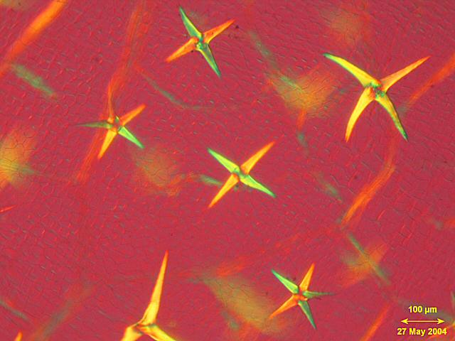
Stellate trichomes (hairs) on leaf of Deutzia gracilis Projectina microscope |
|
|
Transverse section of larch leaf |
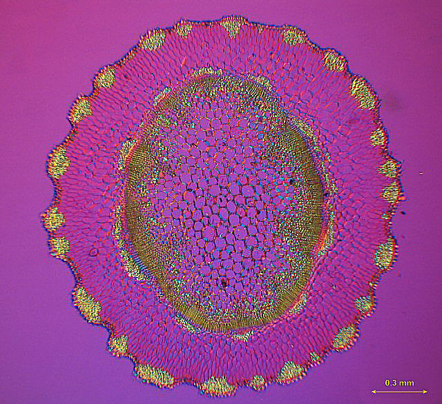
Transverse section of sunflower leaf, viewed in polarised light
|
|
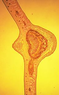
Transverse section of plant stem (Sequiselum spp(?)) Projectina microscope
|
|
|
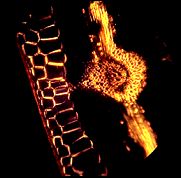
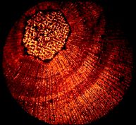
Section of pine twig in polarised light |
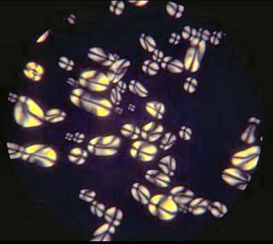
Potato starch in polarised lightTo find more about starch, click on the picture |
|
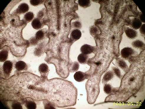
Section through poppy ovaryshowing developing seeds. Original mag = 40x. Olympus FH microscope with Aiptek Pocket Cam 3 digital camera. |
|
|
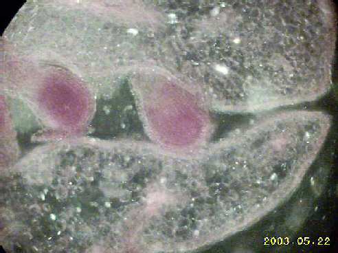
Poppy ovary400x, darkground ilumination (substage wheel-stop) |
|
|
|
|
|
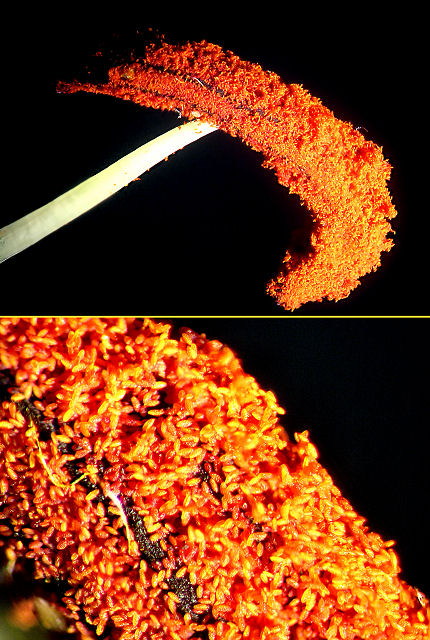
DahliaUpper image: Wild M8 Microscope. Stack of 11 images combined in Helicon Focus Lower image: Wild M8 microscope. Stack of 5 images combined in Helicon Focus |
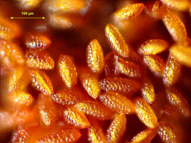
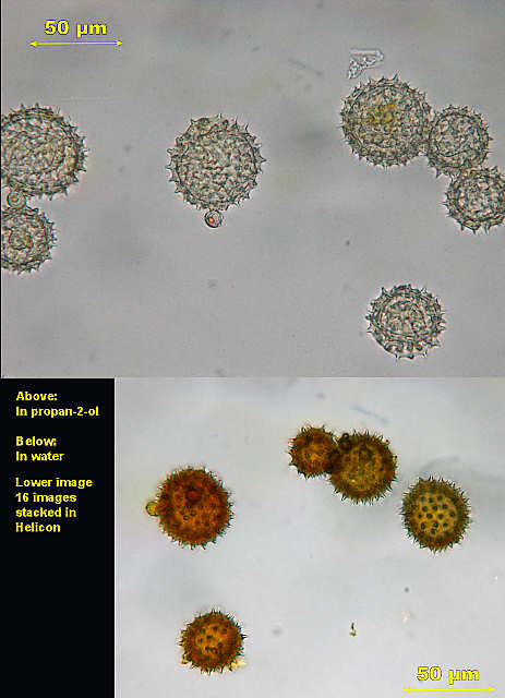
Dahlia PollenBoth images using Zeiss Standard GFL microscope with x40 Zeiss plan acrhomat. The upper image is a single image, the lower a stack of 16 images combined using Helicon Focus. The mountants used were different, as noted above. |
|
HOME: RELOAD MICRO PAGESHOME: RELOAD GARDEN PAGESHOME: RELOAD FRAMES FOR MAIN SITE
|
|